Urgent Appointment
Telehealth
Hornsby Consulting
Bega Operating and Consulting
Canberra Consulting and Operating
Wahroonga
Westmead

For Patients: Urgent Appointment - Message here or Call (02) 9467 5400
For Referrers: 1. Upload referral here
2. Healthlink EDI: ang7sana
3. Argus: argusdocs@specialistsurgeon.com.au
4. Medical Objects

Melanoma
Why is Melanoma 'Australia's national cancer’?
- highest melanoma rates in the world, 16,000 people diagnosed this year.
- most dangerous skin cancer,1300 people will die due to melanoma this year.
- most common cancer affecting 15 to 39-year-old Australians.
- third most common cancer in Australian men, after prostate and colorectal cancer.
- third most common cancer in Australian women, after breast and colorectal cancer
What is Melanoma?
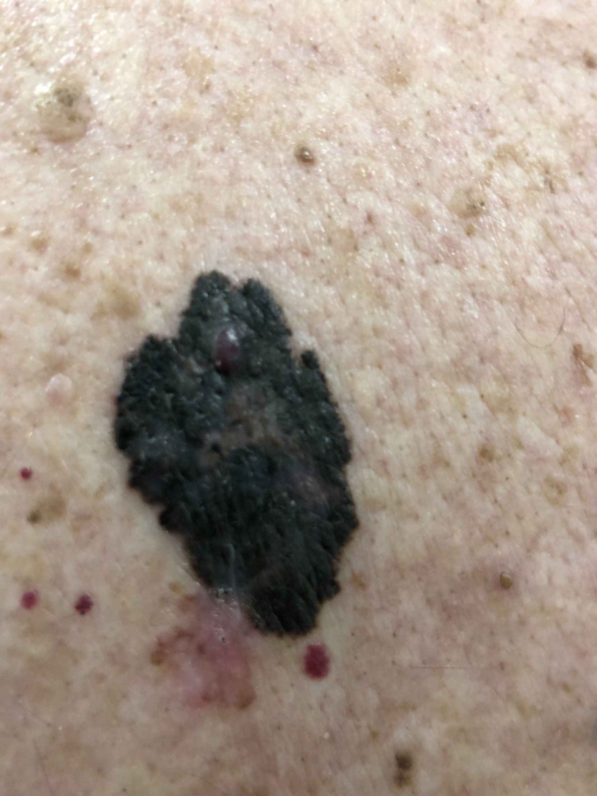
Melanoma is the most dangerous form of skin cancer, these cancerous growths develop when unrepaired DNA damage to skin cells triggers mutations (genetic defects) that lead the skin cells to multiply rapidly and form malignant tumors.
These tumours originate in the pigment-producing melanocytes in the basal layer of the epidermis. Melanocytes function is to produce melanin which protect the skin from ultraviolet (UV) radiation. Melanomas often resemble moles; some develop from moles. The majority of melanomas are black or brown, but they can also be skin-colored, pink, red, purple, blue or white. Here is a
video from the Melanoma Institute of Australia with a comprehensive explanation.
Melanoma is a serious form of skin cancer, it grows quickly and can spread to
- deeper layer of skin
- lymphatic system to spread to lymph glands
- bloodstream and go to other parts of the body e.g. lungs, liver, brain or bone.
What are the Causes of Melanoma?
Melanoma is caused mainly by intense, occasional UV exposure (frequently leading to sunburn), especially in those who are genetically predisposed to the disease.
Extensive exposure to UV radiation from the sun or solarium tanning machines is the most crucial preventable cause. Risk factors include people with fair skin, a high mole count, family history and sunburns throughout life. Other risk factors are a weakened immune system, being older, male and a disorder called Xeroderma Pigmentosum.
It can occur anywhere on the skin, even in hidden areas like soles of feet inside the mouth.
If melanoma is recognized and treated early, it is almost always curable, but if it is not, the cancer can advance and spread to other parts of the body, where it becomes hard to treat and can be fatal.
How to prevent Melanoma and detect it early?
We manage the risk factors, and ‘Sun Safety’ is the single most important practice we can do as a family. Ultra violet (UV) light causes 95% of melanoma.
How do we achieve this?
- avoid exposure to the sun during midday
- protect skin with sunscreen, clothing and a hat when outdoors (even on cloudy days)
- read more on Sun Safety Slip, Slop, Slap, Seek, Slide
If detected early, melanoma can be effectively treated
- new moles and/or existing moles with a recent change must be checked
- regular skin checks at least once every 12 months
What are stages of melanoma ?
The Stage tells us ‘how bad the melanoma is’ which includes a risk profile and an assessment of how much the melanoma has spread. The stages depend on variables noted on a biopsy
- tumour thickness,
- ulceration and the
- involvement of lymph nodes or organs.
Once diagnosed, the stage of melanoma then guides the treatment approach.
What are the ways to stage a melanoma?
- Microstaging - Pathologist examines the specimen under a microscope
- Clinical staging - Lymph node groups draining the melanoma are checked to for any evidence of spread
- Staging after investigation - Scans may be needed CT, MRI, and PET
How is Melanoma diagnosed?
Often, it is diagnosed in a doctor’s office – after patients notice a new mole on their skin or a changes to an existing mole.
Initial Diagnosis
See your doctor as soon as you can, if you see a new dark spot (mole) on your skin or an old spot that’s started changing. A diagnosis that is made early can create a hug difference in the results. Here is what to look for when you do a self examination described in this Melanoma Patients site.
- A Asymmetry
- B Border Irregularities
- C Colour
- D Diameter
- E Evolving
Your doctor will perform a complete head to toe check, a physical examination to check all parts of skin including hands, feet and scalp, also check inside the mouth and nose. A magnifying instrument called a dermoscope may be used to help in the diagnosis. You might be referred to a dermatologist for a further assessment, or you may need a biopsy.
What is a Biopsy?
The recommended method is an excision biopsy, where the whole lesion is removed, but in some cases other techniques may be preferred by your doctor.
An excision biopsy is a small procedure that can be done by your GP specialist, your dermatologist or a surgeon. Starts with a local anaesthetic to numb the region, a scalpel is used to remove the spot along with a small area of surrounding skin. The wound may be closed with few stitches and a small dressing and removed mole is sent to pathology for examination.
What does the Pathologist do?
The pathologist will use a microscope to see if it contains cancer cells and determine the type of cancer and certain features. A review appointment to discuss the results will be made in about a week. You then discuss the tumour progression, it’s depth and risk of spreading. Talking about this process with friends and family will help allay the anxiety around this wait. If cancer cells are found, and melanoma is confirmed, it is staged to see if it has spread which may include taking more skin, i.e. another procedure.
What is a multi disciplinary meeting?
Multidisciplinary teams is the best practice in the treatment planning and care for patients with cancer. It is an integrated team approach to health care in which all members of the oncology team and allied health care professionals meet. All the investigation details, the presentation, pathology and imaging along with patient preferences are discussed. They consider all relevant management options and collaboratively develop a treatment and care plan for each individual patient.
What is the role of Surgery and the Surgical Oncologist?
The most likely and initial treatment for melanoma, and the extent and role depends on the progress of the cancer.
- Early stage melanoma, a biopsy followed by wide local excision is required, where the skin surrounding the melanoma is removed. This is to minimise the risk of recurrence of the melanoma locally.
- Later stage melanoma, surgery is a diagnostic tool to assess the spread of the melanoma. You may need invasive surgery to remove lymph nodes.
Wide Local Excision
The first biopsy usually removes all the visible melanoma, wide local excision is recommended to minimise the risk of the melanoma coming back.
We remove around a 1 cm margin around the site of the original lesion, however this number changes depending on the depth of the melanoma. Primary Closure is usually used, where stitches are used to close the wound, however larger excisions may require skin grafts or skin flaps for reconstruction.
Sentinel Node Biopsy
This operation is performed when a more risky initial (primary) melanoma has been diagnosed by the biopsy in order to see if the melanoma has spread to the regional lymph nodes. This allows us to estimate the risk of the melanoma spreading to other parts of the body.
The procedure involves a preoperative lymphatic mapping the day before or the morning of the surgery. Radioactive dye is injected into the skin around the melanoma, where it drains into the lymphatic channels. Lymphatic mapping identifies the first gland to receive (drain) the dye, this is called the sentinel node, there may be one, two sometimes three.
It is removed and sent to pathology, where after an examination under the microscope, malignant cells if present are detected. Since this is an indicator of the risk that melanoma may have spread to other parts of the body, other imaging tests like CT, MRI and/or PET scans may be appropriate.
Lymph Node Dissection
Melanoma that has spread to sentinel nodes in the draining node groups in the armpit, neck or groin may be confined to these regions before spreading to other organs. This may be treated with surgery called regional lymph node clearance that removes all the lymph nodes in the area. There are three types of regional lymph node clearance: axillary (armpit), neck and groin.
Lymph node dissection removes all the lymph glands and possible cancer containing tissue. It is done under a general anaesthetic so you will not be aware of the procedure. Local anaesthetic is injected into the site of surgery for pain relief after the operation.
There are several important arteries, veins and nerves that are present in these areas. The operation is performed to remove the lymph nodes and cancer tissue without causing damage (if possible) to any important structures.
The procedure takes 60 to 90 minutes and a surgical drain is inserted to collect the fluid produced postoperatively while the wound heals. The silicone drain is a flexible soft tube connected to a suction bottle and will be monitored by nursing staff, kept in for several days to weeks after the procedure.
The tissue that has been removed is sent to pathology, and again there is a wait of around a week for the final results.
Recovery from these procedures requires 2 - 7 days of bed rest after your operation. Analgesia will be provided to manage post-operative pain and you will be given instructions on dressings and activity after surgery.
What is Lymphoedema?
This is a common side effect of lymph node dissection surgery. Blockage in the lymphatic system after surgery causes swelling of limbs, may feel tight and may be difficult to use.
The frequency and severity depends on certain factors
- the extent of surgery (or radiotherapy)
- body weight before treatment
- natural resistance to lymphedema.
Early treatment can manage and reverse the swelling. Minimal swelling may respond to compression therapy, and massage and exercise may help get the fluid moving. It is important to have good skin care to keep skin healthy.
Reconstructive Surgery
An important sequelae of melanoma surgery is scarring of skin or altered function e.g. damaged nerves or loss of movement.
Reconstructive surgery offers an opportunity to manage these problems and help patients live their best lives.
Skin grafts – used when the Wide Local Excision leaves a wound area too big for stitches. Skin is taken from another part of the body and used to reconstruct the wound.
Flap repairs – skin close to the defect is lifted and moved over the wound, providing an excellent match in colour and texture.
Radiation therapy
This treatment uses x-rays to kill cancer cells by damaging their DNA and since cancer cells cannot repair themselves, they die. They are broken down and eliminated by the body.
The treatment is carefully planned to allow the normal cells to repair themselves and minimise side effects. The total dose and the number of treatments you need depends on the size and location of the melanoma, your general health and other concurrent medical treatments.
This radiation can come from a radiation machine or radioactive material placed in your body near the cancer.
Side effects of this treatment can be
- Temporary or permanent loss of hair in the area
- Skin irritation
- Temporary change in skin colour
- General fatigue
- Other side effects depending on the area of the body
What are targeted therapies?
This is a drug that blocks the growth of cancer by interfering with specific molecules involved in tumour growth, used for patients with the spread of the disease to other organs in the body.
They target some of the key genetic mutations that drive the growth of melanoma in patients and selectively block activity of these driving mutations.
Genetic mutations involved in melanoma development so far are:
- BRAF
- NRAS
- C-KIT
What is immunotherapy?
These are treatments that work to trigger the body’s immune system to fight the cancer cells. The methods are – activating an immune response with checkpoint inhibitors or “training” an immune response with vaccines.
How do immunotherapies work?
Checkpoint inhibitors, drugs stimulate the immune system to recognise and destroy melanoma cells.
Immunotherapy currently available
- Ipilimumab
- Pembrolizumab
- Nivolumab
Side Effects – most common side effects of immunotherapy include:
- skin problems (redness, dry skin, rash, blistering)
- flu-like symptoms (feeling tired, fever, weakness)
- diarrhoea
- cough
- breathing difficulty
- neurological problems (weakness/numbness)
What is the role of Chemotherapy in Melanoma?
Anti-cancer drugs slow the growth of tumour cells that have spread to internal organs and may help relieve symptoms caused by the growth of the tumour.
New targeted therapies focused on the genes of melanoma cells are effective treatment but in some rare cases chemotherapy may be recommended.
Side effects
- nausea, vomiting
- depression
- tiredness
- hair loss
These side effects are temporary and steps can be taken to prevent or reduce them.
cancers are almost always removed. In more advanced skin cancers, some of the surrounding tissue may also be removed to make sure that all of the cancerous cells have been taken out.
There are many treatment options available depending on the stage of melanoma.
Localised (early stage) melanoma needs surgery, and in the majority of cases, may be the only treatment required.
More advanced cases of melanoma where the cancer has spread to lymph glands or other parts of the body may require treatments such as chemotherapy, radiotherapy, immunotherapy or targeted molecular therapy. This involves first multiple imaging modalities to determine the extent of the condition.
Your doctor will recommend the best treatment option based on how far the melanoma has progressed together with other factors such as your age and general health. This will be discussed at a multidisciplinary meeting, to get the best possible individualised treatment.
What if Melanoma is Untreated?
While it is not the most common of the skin cancers, it causes the most deaths. Melanoma kills about one in six patients.
For Patients
For Referrers
Quick Enquiry
Thank you for contacting us. We will get back to you as soon as possible.
Oops, there was an error sending your message. Please try again later.
IMPORTANT PAGE LINKS
PRACTICE DETAILS
Wahroonga Specialist Centre
Suite 9/176 Fox Valley Rd, Wahroonga, NSW, 2076
Westmead Private Hospital
Suite 212, 12 Mons Rd, Cnr Darcy Rd, Westmead, NSW 2145
Calvary Bruce Private Hospital (Operating)
30 Mary Potter Cct, Bruce, ACT 2617
OPEN HOURS
- Mon - Fri
- -
- Saturday
- Appointment Only
- Sunday
- Closed
IMPORTANT PAGE LINKS
PRACTICE DETAILS
OPEN HOURS
- Mon - Fri
- -
- Saturday
- Appointment Only
- Sunday
- Closed
All Rights Reserved | Dr Sandra Krishnan


